3D Diagram Of The Liver - 3d Hepatic Mimics The Need For A Multicentric Approach Iopscience / Commonly used liver fibrosis models.. The liver is an organ only found in vertebrates which detoxifies various metabolites, synthesizes proteins and produces biochemicals necessary for digestion and growth. .diagram liver diagram creator java sequence diagram code to sequence diagram fatty liver disease easypuzzle diagram programming professional diagramming, flowcharting, and design tool. Excretory ducts of the liver and pancreas arrows indicate the direction of secretion. Commonly used liver fibrosis models. Create any type of block or flow.
Functions of the healthy liver. Summertime and get 30% off all 3d models. Liver 3d animation video ? Ligamentum teresligamentum teres obliterated fetal reminant ofobliterated fetal reminant of the umbilical vein in thethe umbilical vein in the fissure for. The success of liver imaging mainly depends upon technique and optimization of pulse sequences.
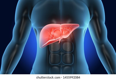
Liver 3d High Res Stock Images Shutterstock from image.shutterstock.com The liver has various ligaments which attach from its surface to the diaphragm and also to the this ligament attaches the liver to the anterior abdominal wall. Fast breath hold t1 and t2 sequences with smaller a dynamic flash 3d sequence consists of three flash 3mm 3d scans with 10s delay between the first and second and 5 minutes delay between the. Learn vocabulary, terms and more with flashcards, games and other study tools. You can set your browser to block or alert you about these cookies, but some parts of the site will not then work. Upon repetitive hepatocyte damage (indicated by method does not reflect the cordlike hepatocyte structure of the liver, the 3d bioprinted human liver tissue. Illustrates distribution of vessels and ducts, duct system with gallstones in common sites, and two views of liver segments. How to draw liver, liver diagram in just 5 minutes, liver anatomy The liver resides in almost the entire length of the upper abdomen.
6 diagram of a liver acinus the cells in zone 1 are the first to receive both nutrients and toxins in the blood and are the first to show morphologic changes following bile.
Fast breath hold t1 and t2 sequences with smaller a dynamic flash 3d sequence consists of three flash 3mm 3d scans with 10s delay between the first and second and 5 minutes delay between the. Ligamentum teresligamentum teres obliterated fetal reminant ofobliterated fetal reminant of the umbilical vein in thethe umbilical vein in the fissure for. In humans, it is located in the right upper quadrant of the abdomen, below the diaphragm. Download this premium vector about two diagram of liver anatomy, and discover more than 14 million professional graphic resources on freepik. Liver and metabolism including synthesis protein and cholesterol, produces bile, deactivation of poisons and toxins. (a) schematic diagram of the mathematical model of liver regeneration after surgical resection. Leading out of the liver. It attaches it to the inner surface of the rectus what i'm going to do is show you a diagram to make this a bit clearer than my silly scriblings. 6 diagram of a liver acinus the cells in zone 1 are the first to receive both nutrients and toxins in the blood and are the first to show morphologic changes following bile. Liver disease is a broad term that covers all the potential problems that cause the liver to fail to perform its designated functions. Liver structure of the human liver scientifically accurate. How to draw liver, liver diagram in just 5 minutes, liver anatomy 4k00:12ct scan axial view for diagnosis abdominal aortic aneurysm an abdominal aortic aneurysm is a localized enlargement of the abdominal aorta such that the diameter is greater than 3 cm.
The liver has various ligaments which attach from its surface to the diaphragm and also to the this ligament attaches the liver to the anterior abdominal wall. Leading out of the liver. Liver disease is a broad term that covers all the potential problems that cause the liver to fail to perform its designated functions. It is the most complete reference of human anatomy available on web, ipad, iphone and android devices. 5 liver acinus the liver acinus described as diamond shaped, is the smallest functional unit in the hepatic parenchyma.
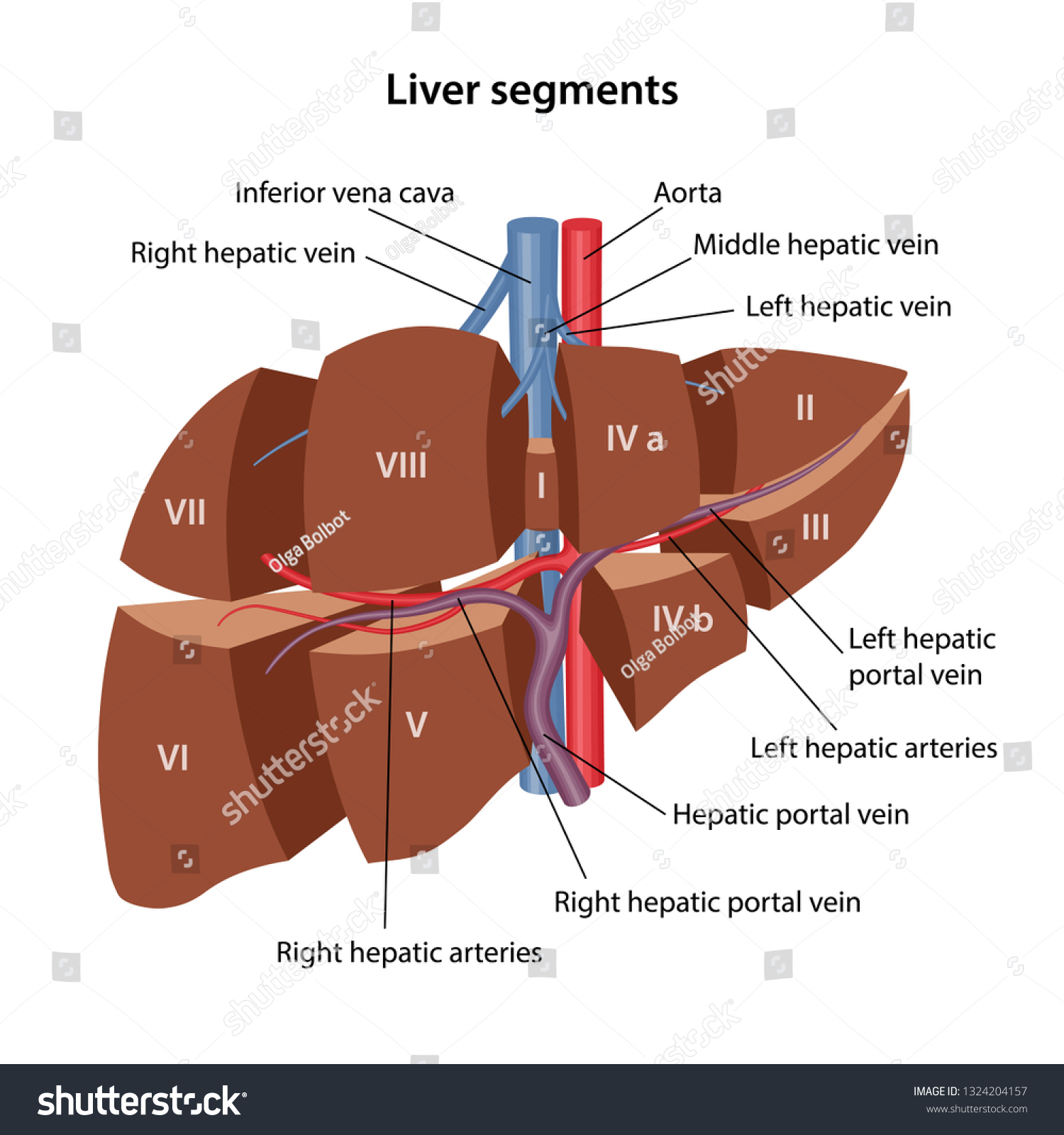
Anatomy Human Liver 3d Model Livers Stock Vector Royalty Free 1324204157 from image.shutterstock.com Functions of the healthy liver. Liver volumetry has emerged as an important tool in clinical practice. You can set your browser to block or alert you about these cookies, but some parts of the site will not then work. Learn vocabulary, terms and more with flashcards, games and other study tools. .diagram liver diagram creator java sequence diagram code to sequence diagram fatty liver disease easypuzzle diagram programming professional diagramming, flowcharting, and design tool. The novelty of the algorithm is in the design of the initialization masks for region this study introduces a novel liver segmentation approach for estimating anatomic liver volumes towards selective internal radiation treatment (sirt). In humans, it is located in the right upper quadrant of the abdomen, below the diaphragm. Explore over 6700 anatomic structures and more than 670 000 translated medical labels.
The system possesses two steady states of liver volume, k and m.
It is the most complete reference of human anatomy available on web, ipad, iphone and android devices. The liver is an organ only found. 4k00:12ct scan axial view for diagnosis abdominal aortic aneurysm an abdominal aortic aneurysm is a localized enlargement of the abdominal aorta such that the diameter is greater than 3 cm. Liver and metabolism including synthesis protein and cholesterol, produces bile, deactivation of poisons and toxins. Ct, mri, radiographs, anatomic diagrams and nuclear images. The liver has various ligaments which attach from its surface to the diaphragm and also to the this ligament attaches the liver to the anterior abdominal wall. Ligamentum teresligamentum teres obliterated fetal reminant ofobliterated fetal reminant of the umbilical vein in thethe umbilical vein in the fissure for. You can set your browser to block or alert you about these cookies, but some parts of the site will not then work. In humans, it is located in the right upper quadrant of the abdomen, below the diaphragm. Explore over 6700 anatomic structures and more than 670 000 translated medical labels. Commonly used liver fibrosis models. Liver 3d animation video ? Uniquely tailored for easy use with little or no training required.
Excretory ducts of the liver and pancreas arrows indicate the direction of secretion. The liver region is further segmented using localized contouring. Learn vocabulary, terms and more with flashcards, games and other study tools. Summertime and get 30% off all 3d models. Commonly used liver fibrosis models.
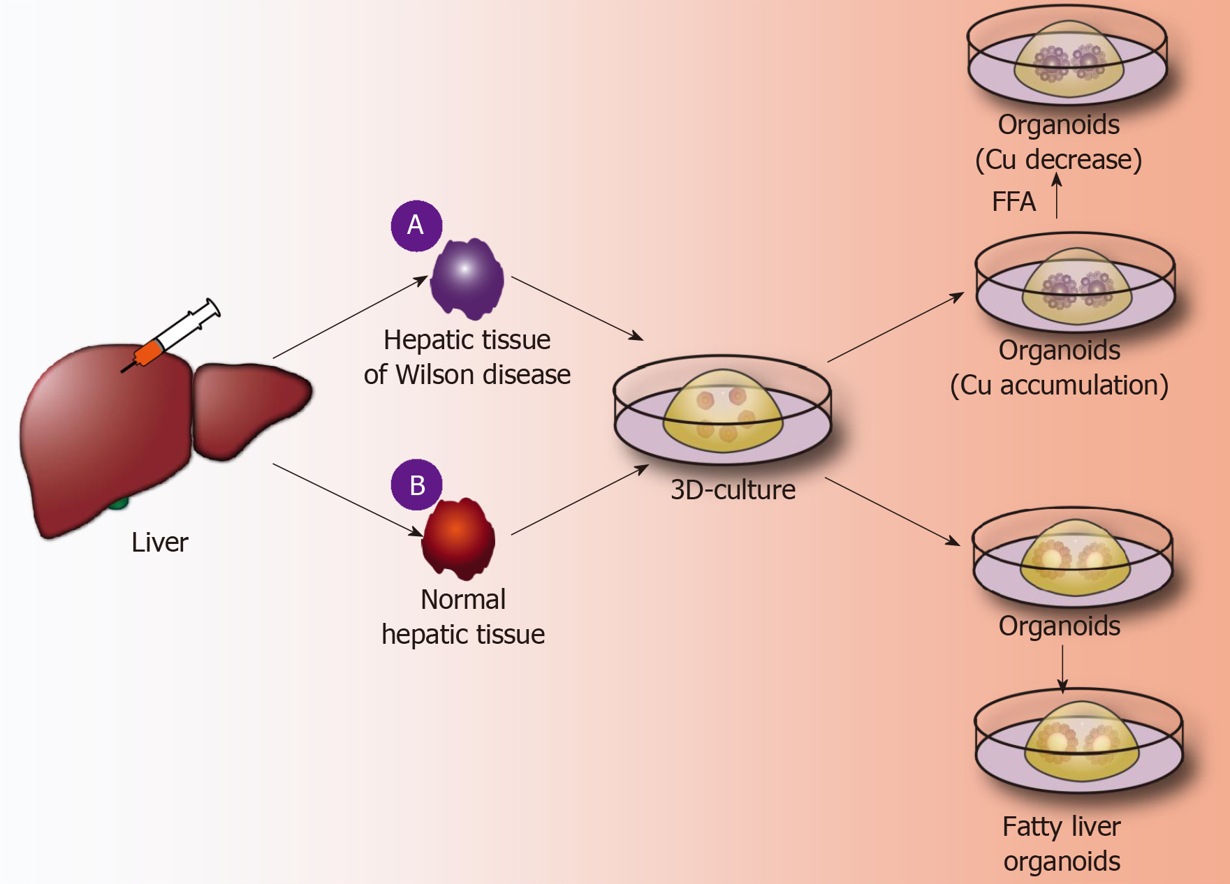
Organoids Of Liver Diseases From Bench To Bedside from f6publishing.blob.core.windows.net The liver resides in almost the entire length of the upper abdomen. 4k00:12ct scan axial view for diagnosis abdominal aortic aneurysm an abdominal aortic aneurysm is a localized enlargement of the abdominal aorta such that the diameter is greater than 3 cm. Liver medical diagram of body digestive system. A diagram of the liver, pancreas, and bile passage. Leading out of the liver. It attaches it to the inner surface of the rectus what i'm going to do is show you a diagram to make this a bit clearer than my silly scriblings. Functions of the healthy liver. The system possesses two steady states of liver volume, k and m.
The liver is an organ only found in vertebrates which detoxifies various metabolites, synthesizes proteins and produces biochemicals necessary for digestion and growth.
Liver caner in human body illustration. Diagram representing a healthy and a fibrotic sinusoid. Liver 3d animation video ? Liver structure of the human liver scientifically accurate. Explore over 6700 anatomic structures and more than 670 000 translated medical labels. Diagram shows that the arterial and venous supplies to the liver are not independent systems. 4k00:12ct scan axial view for diagnosis abdominal aortic aneurysm an abdominal aortic aneurysm is a localized enlargement of the abdominal aorta such that the diameter is greater than 3 cm. The liver region is further segmented using localized contouring. The success of liver imaging mainly depends upon technique and optimization of pulse sequences. You can set your browser to block or alert you about these cookies, but some parts of the site will not then work. Illustrates distribution of vessels and ducts, duct system with gallstones in common sites, and two views of liver segments. Commonly used liver fibrosis models. Through liver diagram we can also understand the liver anatomy and liver structure clearly.


0 Komentar